Assay points.
-
ASBF-PST3-B500
500
-
ASBF-PST3-B10K
10,000
-
ASBF-PST3-B50K
50,000
-
ASBF-PST3-B-HV
100
Assay Principle
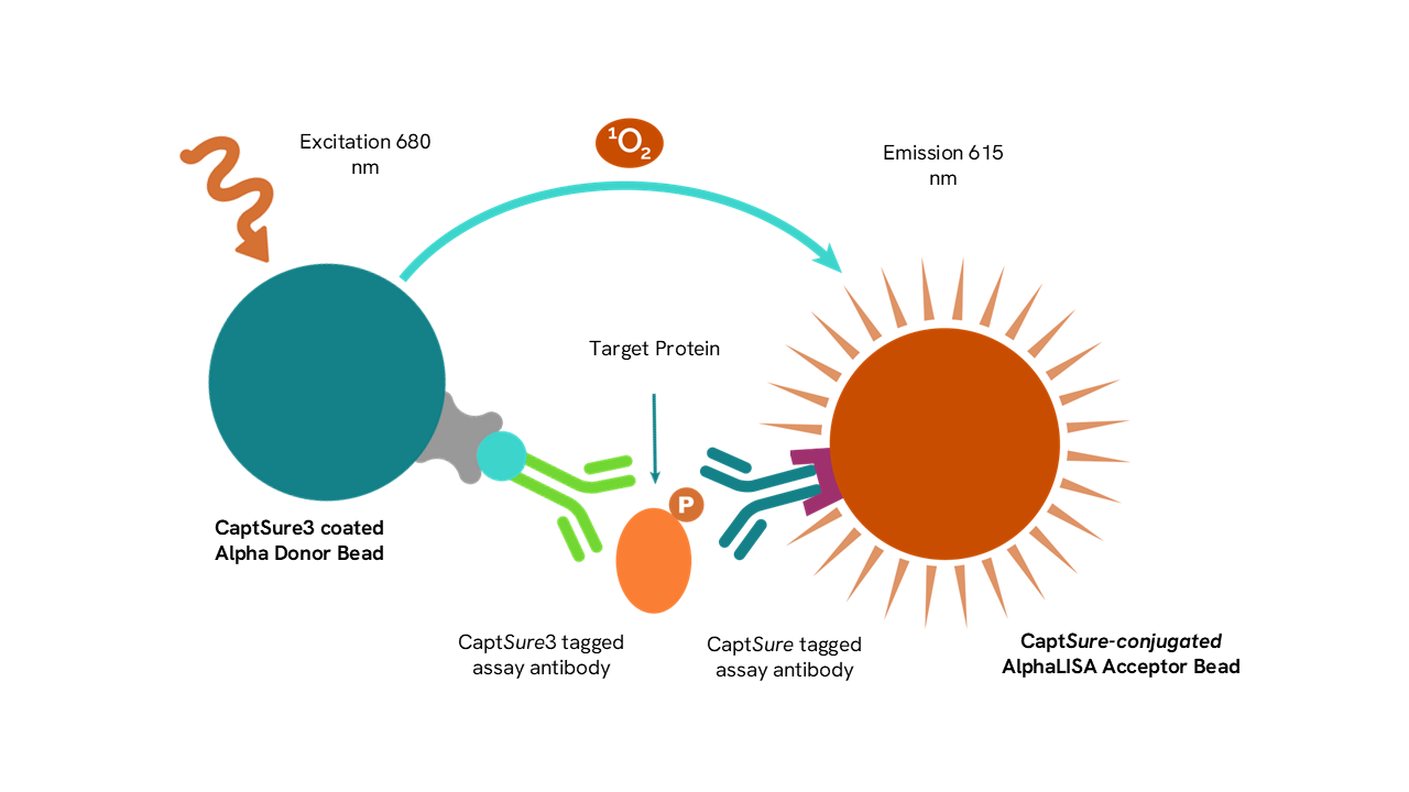
The AlphaLISA™ SureFire® Ultra™ assay enables the rapid and sensitive detection of total and phosphorylated cellular proteins. AlphaLISA™ assays utilize two bead types: Acceptor Beads and Donor Beads. The Acceptor Bead is coated with a proprietary CaptSure™ agent to specifically immobilize the assay specific antibody which is labeled with a CaptSure™ tag. The Donor Bead is coated with streptavidin to capture the biotinylated antibody.
In the presence of a target protein, the two target-specific antibodies bring Donor and Acceptor Beads into close proximity. When the Donor Beads are activated by a laser (680 nm), singlet oxygen is transferred to the Acceptor Bead leading to the production of an Alpha signal. The amount of light emission (615 nm) from the Acceptor Bead is directly proportional to the amount of target protein present in the sample. If an Acceptor Bead is not in close proximity (i.e. 200 nm) of a Donor bead, little to no signal is produced over background.
The assay can be executed in a 1-plate or 2-plate assay protocol (Refer to Manual for more details).
1-plate assay protocol: culturing of cells, treatment, lysis and assay are performed in a single well, enabling miniaturization in high throughput screening programs.
2-plate assay protocol: cells are cultured and treated in a 96-well culture plate and lysates are then transferred into a separate plate for assay. This format allows the evaluation of multiple targets from a single lysate.
Technical Specific Data
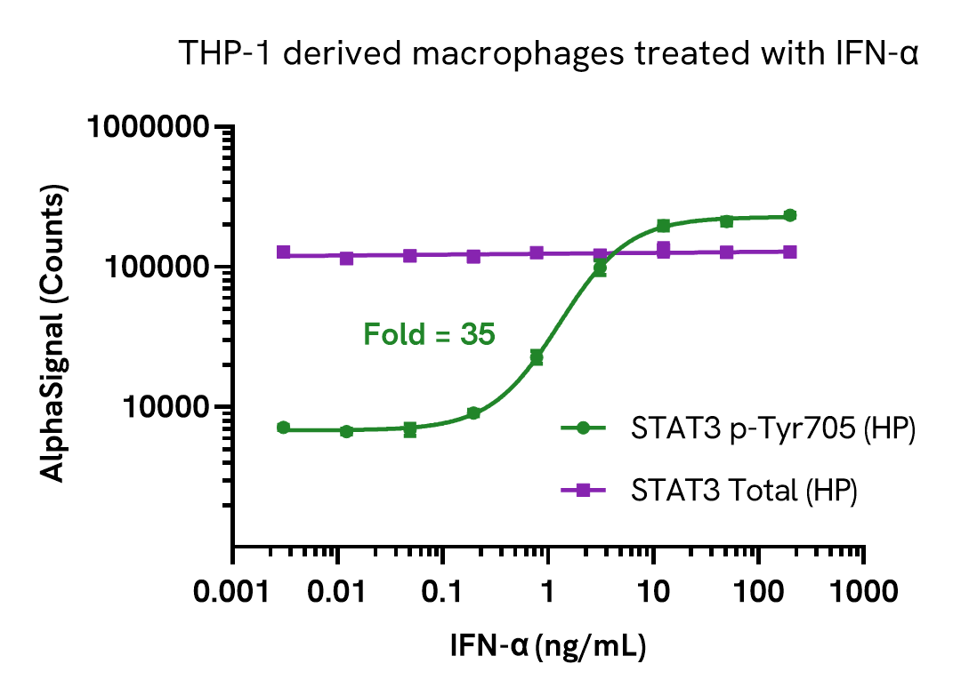
Data obtained with a 2-plate, 2-incubation protocol. THP-1 cells were seeded in a 96-well plate (50,000 cells/well) in complete medium containing 100 nM PMA and incubated for 24 hours. The THP-1 derived macrophages were serum starved for 2 hours and then stimulated with increasing concentrations of IFNα for 20 minutes. After treatment, the cells were lysed with Lysis Buffer and assayed separately for Phospho (Tyr705) and Total STAT3 using respective SureFire® Biotin Free High Performance kits. Equivalent to approximately 5,000 cells/datapoint.
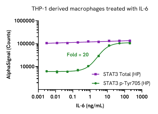
Data obtained with a 2-plate, 2-incubation protocol. THP-1 cells were seeded in a 96-well plate (50,000 cells/well) in complete medium containing 100 nM PMA and incubated for 24 hours. The THP-1 derived macrophages were serum starved for 2 hours and then stimulated with increasing concentrations of IL-6 for 30 minutes. After treatment, the cells were lysed with Lysis Buffer and assayed separately for Phospho (Tyr705) and Total STAT3 using respective SureFire® Biotin Free High Performance kits. Equivalent to approximately 5,000 cells/datapoint
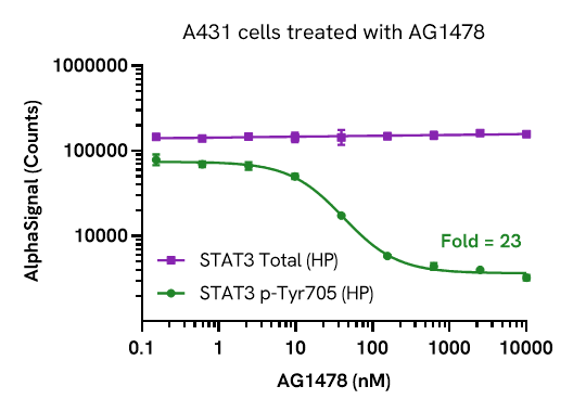
Data obtained with a 2-plate, 2-incubation protocol. A431 cells were seeded in a 96-well plate (40,000 cells/well) in complete medium and incubated overnight. Cells were then treated with increasing concentrations of AG1478 for 2 hours and then stimulated with a fixed 100 ng/mL of EGF for 15 minutes. After treatment, the cells were lysed with Lysis Buffer and assayed separately for Phospho (Tyr705) and Total STAT3 using respective SureFire® Biotin Free High Performance kits. Equivalent to approximately 4,000 cells/datapoint.
Manuals & downloads.
-
PDF
-
PDF
Detection Kit Manual
-
Tech Spec
Technical Data Sheet
Safety Data Sheets
-
Safety Data Sheet (SDS)
Safety Data Sheet EU-EN
-
Safety Data Sheet (SDS)
Safety Data Sheet US